Osteochondritis Dissecans | |
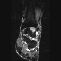
Osteochondritis dissecans of the talus is typically seen in young, athletic individuals, and results from trauma. The process represents a fracture across the articular surface of the talus. The fracture fragment may migrate into the joint forming a loose body. Radiographically, the lesion is seen as a well circumscribed lucency with a sclerotic rim. MRI shows edema surrounding the lesion. The classic location for osteochondritis dissecans is the lateral aspect of the medial condyle of the femur, but may be seen elsewhere including the femoral head, radial head, talus, and posterior patella.
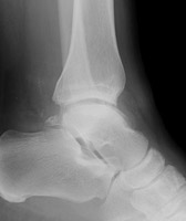 A A | Lateral radiograph of the ankle. The abnormality is not seen. |
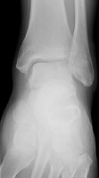 B B | Mortise radiograph of the ankle. There is a radiolucent lesion at the lateral aspect of the talus, corresponding to the site of osteochondritis dissicans. |
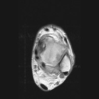 C C | Axial MRI. This proton density image of the ankle demonstrates a region of intermediate signal intensity at the lateral aspect, corresponding to edema. |
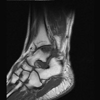 D D | Sagittal MRI. This T1-weighted image shows a region of low signal intensity representing the site of osteochondritis dissecans and edema. |
 E E | STIR image of the ankle. The region of intermediate signal intensity on the PD image here is seen as bright signal, corresponding to edema. |
Links to online textbooks:
Links to online cases:
References:
 A
A B
B C
C D
D E
E