![]()
![]()
![]()
![]()
![]()
![]()
Atlas of Signs in Musculoskeletal Radiology is approved by the ARRS (American Roentgen Ray Society) and is included in AJR Webreview
A. Gentili,MD, M. Beller, MD, S. Masih, MD, L.L. Seeger, MD
![]()
|
Atlas of Signs in Musculoskeletal Radiology is approved by the ARRS (American Roentgen Ray Society) and is included in AJR WebreviewA. Gentili,MD, M. Beller, MD, S. Masih, MD, L.L. Seeger, MD
|
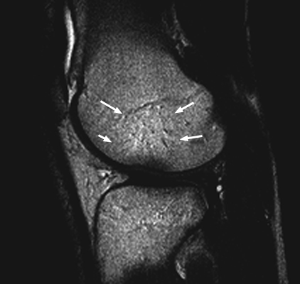
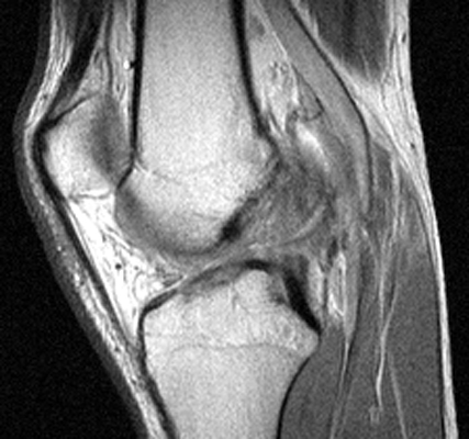
Diagnosis:ACL Tear Discussion:Sagittal T2 weighted and proton density images which reveal complete tear of the ACL with mild increase in marrow signal in the adjacent osseous structures. The bone bruises, as evidenced by increased signal within the marrow, is likely caused by anterior subluxation of the tibia at the time of tear of the ACL accompanied by impaction of the middle portion of the lateral femoral condyle against the posterior portion of the lateral tibial plateau. Signal intensity abnormalities are probably secondary to edema, hemorrhage, and microfracture.
References:
|
|
Atlas of Signs in Musculoskeletal Radiology is approved by the ARRS (American Roentgen Ray Society) and is included in AJR WebreviewA. Gentili,MD, M. Beller, MD, S. Masih, MD, L.L. Seeger, MD
|