![]()
![]()
![]()
![]()
![]()
![]()
Atlas of Signs in Musculoskeletal Radiology is approved by the ARRS (American Roentgen Ray Society) and is included in AJR Webreview
A. Gentili,MD, M. Beller, MD, S. Masih, MD, L.L. Seeger, MD
![]()
|
Atlas of Signs in Musculoskeletal Radiology is approved by the ARRS (American Roentgen Ray Society) and is included in AJR WebreviewA. Gentili,MD, M. Beller, MD, S. Masih, MD, L.L. Seeger, MD
|
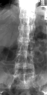
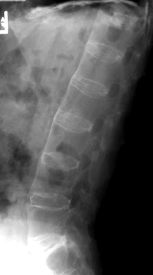
Diagnosis:Ankylosing Spondylitis Discussion:Anterior and lateral radiograph of the lumbar spine which reveals near complete fusion of all of the vertebral bodies. Spinal changes which include squaring of the vertebral bodies and fusion between adjacent levels. The fusion is secondary to syndesmophytes and resembles a bamboo stalk. This is most commonly found in ankylosing spondylitis.
References:
|
|
Atlas of Signs in Musculoskeletal Radiology is approved by the ARRS (American Roentgen Ray Society) and is included in AJR WebreviewA. Gentili,MD, M. Beller, MD, S. Masih, MD, L.L. Seeger, MD
|