![]()
![]()
![]()
![]()
![]()
![]()
Atlas of Signs in Musculoskeletal Radiology is approved by the ARRS (American Roentgen Ray Society) and is included in AJR Webreview
A. Gentili,MD, M. Beller, MD, S. Masih, MD, L.L. Seeger, MD
![]()
|
Atlas of Signs in Musculoskeletal Radiology is approved by the ARRS (American Roentgen Ray Society) and is included in AJR WebreviewA. Gentili,MD, M. Beller, MD, S. Masih, MD, L.L. Seeger, MD
|
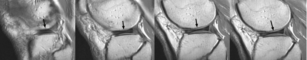
Diagnosis:Discoid Meniscus Discussion:Seen on sagittal images, the bow-tie represents the continuity of the meniscus between the anterior and posterior horns. It is normally seen on 2 contiguous sagittal images. When it is seen on 3 or more contiguous 5 mm thick sagittal images is indicative of a discoid meniscus.
References:
|
|
Atlas of Signs in Musculoskeletal Radiology is approved by the ARRS (American Roentgen Ray Society) and is included in AJR WebreviewA. Gentili,MD, M. Beller, MD, S. Masih, MD, L.L. Seeger, MD
|