![]()
![]()
![]()
![]()
![]()
![]()
![]()
![]()
![]()
|
Advanced Imaging of the Diabetic Foot and Its
Complications
|
|
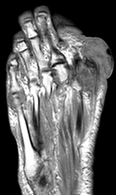
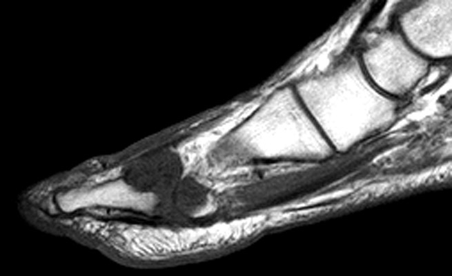
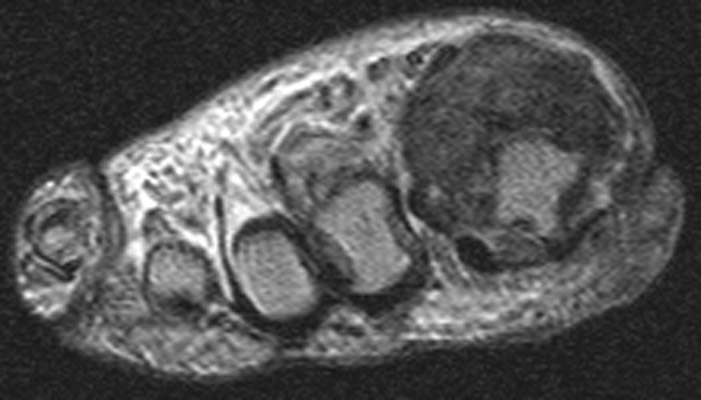
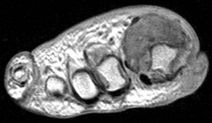
77-year-old man with gout. A. Sagittal T1 weighted, B. Long axis proton density, C. short-axis T2 weighted and D. short-axis proton density images of the right foot demonstrate a large mass with intermediate to low signal intensity on all sequences and underlying erosiond of the 1st metatarsal head in a patient with tophaceus gout. (Click on the images to see larger versions)
|
|
Advanced Imaging of the Diabetic Foot and Its
Complications
>
|