![]()
![]()
![]()
![]()
![]()
![]()
![]()
![]()
![]()
|
Advanced Imaging of the Diabetic Foot and Its
Complications
|
|
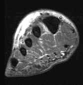
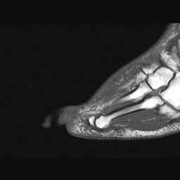
Edema and inflammation of soft tissues is the most common MR imaging finding in the diabetic foot demonstrated by increased signal changes seen on T2 and STIR sequences. Areas of soft tissue edematous change are frequently seen in the plantar forefoot just deep to the plantar aponeurosis and in the dorsum of the foot. These findings do not always correlate with inflammation and cellulitis and may be incidentally seen in patients who do not have clinical suspicion of infection in these areas. The likely cause of these findings may be nonspecific edematous change and cardiogenic in nature. The identification of cellulitic change however remains important as theses areas do need to be scrutinized carefully for foreign bodies, ulcers, abscesses, and osteomyelitis.
Advanced Imaging of the Diabetic Foot and Its Complications
|
|
Advanced Imaging of the Diabetic Foot and Its
Complications
>
|
 A
A
 B
B