![]()
![]()
![]()
![]()
![]()
![]()
![]()
![]()
![]()
|
Advanced Imaging of the Diabetic Foot and Its
Complications
|
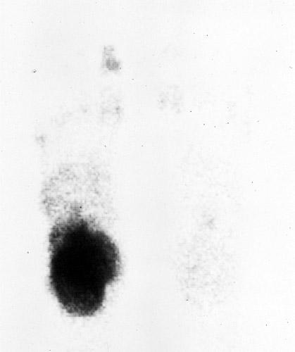
ScintigraphyThree Phase Bone scanThe first phase or angiographic phase is obtained at the time of administration of the radiopharmaceutical (Tc-99m-MDP), the second phase or blood pool phase is obtained in the first few minutes after injection, and the third phase is obtained 3-6 hours after injection. In soft-tissue infections the first two phases demonstrate increased uptake, but the third phase is normal or demonstrates diffuse mildly increased uptake. In osteomyelitis all three phases demonstrate increased uptake, and usually the uptake in the third phase is more focal. However, in neuropathic arthropathy increased uptake can be present in all three phases, for this reason three-phase bone scans, although have a high sensitivity have a low specificity for osteomyelitis in the diabetic foot. To improve the specificity a 24-hr image to create a four-phase bone scan has been advocated. If the lesion is osteomyelitis the amount of uptake in the lesion versus the amount in the normal bone continues to increase-. False negative exams can be caused by ischemia; the radiotracer must be able to reach the foot to accumulate in the focus of osteomyelitis.
Table 2. Three Phase Bone Scan in Osteomyelitis
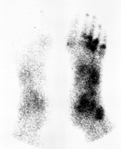
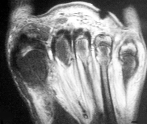
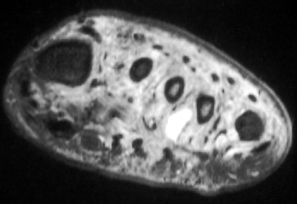
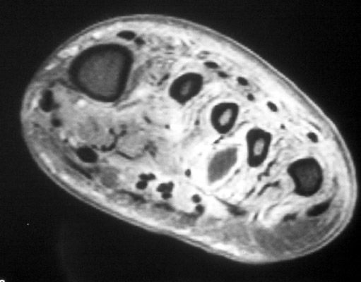
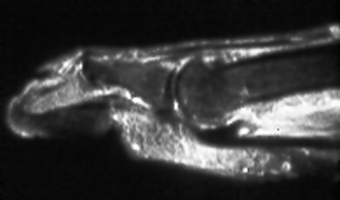
55-year-old man with diabetes. (A) Nuclear medicine bone scan demonstrates abnormal tracer activity in the forefoot consistent with osteomyelitis. (B-C) Long and short axis STIR images of the forefoot demonstrate multiple areas of abnormal bone marrow signal consistent with multifocal osteomyelitis as well as abscess in the plantar soft tissues. (D) Short axis T1 weighted image with fat saturation post intravenous gadolinium administration demonstrates typical enhancement of the soft tissues peripheral to the abscess. (Click on the images to see larger versions)
Gallium-67 ScintigraphyGallium-67 citrate localizes in areas of osteomyelitis by granulocyte and bacterial uptake, and by binding to lactoferrin at the site of infection. However, Gallium-67 citrate is also a bone agent and accumulates at site of increased bone remodeling as in neuropatic joints. Gallium scans are interpreted in conjunction with bone scans, if the distribution of the gallium uptake is different from the uptake on the bone scan, it represents osteomyelitis. In direct comparisons labeled white blood cell scans are superior to gallium scans, and gallium scans is now seldom used for detection of osteomyelitis, if labeled white blood cell scans are available.
Labeled White Blood CellsLabeled leukocytes accumulate in areas of infection, and are more specific for infection then gallium, especially when infection is superimposed on different process causing bone remodeling. Leukocytes have been labeled for many years with indium-111, and more recently with technetium-99m-HMPAO. The advantages of using technetium labeled leukocytes is that technetium is readily available and a larger dose can be use improving image quality. Due to poor spatial resolution of indium labeled white blood cells can be difficult to differentiate infection in bone from infection in the adjacent soft tissues. Using combined bone scan and indium labeled leukocytes is easier to determine if the infection is in soft tissue or bone. However, findings of rapidly progressing, noninfected neuropatic osteoarthropathy on bone scans, indium labeled leukocytes and MRI can be indistinguishable from those of osteomyelitis.
Table 3. In-111 Labeled Leukocytes in Osteomyelitis
| ||||||||||||||||||||||||||||||||||||||||||||||||||||||||||||||||||||||||||||||||||||||||||||||||||||||||||||||||||
|
Advanced Imaging of the Diabetic Foot and Its
Complications
>
|