![]()
![]()
![]()
![]()
![]()
![]()
![]()
![]()
|
Advanced Imaging of the Diabetic Foot and Its
Complications
|
RadiographsThe fist imaging study should be a plain radiograph. Radiographs are usually obtained in the anteroposterior, oblique and lateral projection. The use of single emulsion, high resolution x-ray film\screen combination facilitates the detection of early changes. Changes indicative of osteomyelitis include cortical erosions, permeative radiolucency, and periosteal new bone formation. However radiographic changes may not be seen for 1 to 2 weeks after the onset of acute osteomyelitis, and often is not easy to distinguish osteomyelitis from neuropatic arthropathy especially in the tarsal and tarso-metatarsal regions. Radiographs may demonstrate gas in the soft tissue. Gas can enter the soft tissue through ulcers, skin lacerations, may be introduced during operative procedure, or may be produced by bacteria such as Escherichia Coli, Streptococci Bacteroides, and Clostridium Perfrigerans.
| ||||||||||||||||||||||||||||||||||||||||||||||||||||||||||||
|
Advanced Imaging of the Diabetic Foot and Its
Complications
>
|
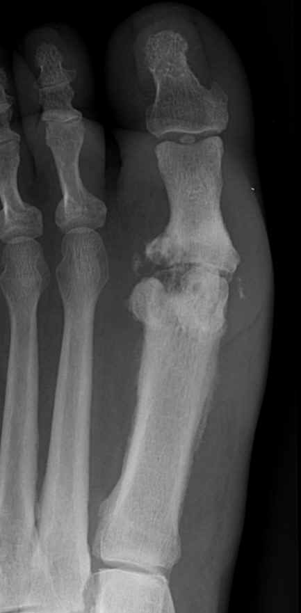 Table 1. Plain
Radiographs in Osteomyelitis
Table 1. Plain
Radiographs in Osteomyelitis