![]()
![]()
![]()
![]()
![]()
![]()
![]()
![]()
![]()
|
Advanced Imaging of the Diabetic Foot and Its
Complications
|
|
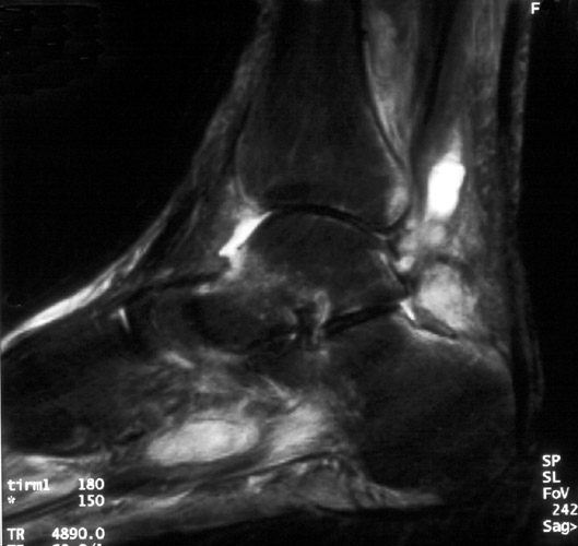
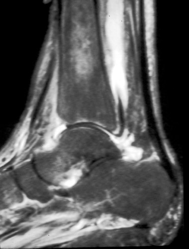
Septic arthritis in the diabetic foot usually arises from contiguous osteomyelitis spreading into the joint. Occasionally septic arthritis may arise from penetrating injury into the joint. Joint space narrowing and destruction ensues, accompanied by joint effusion. The septic joint fluid may communicate with adjacent structures such as tendon sheaths.
 B B
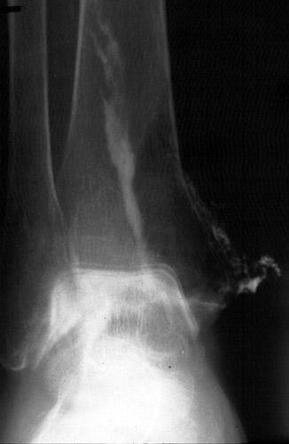
A. Sagittal STIR image of patient with septic tibio-talar joint. The joint fluid communicates with the flexor digitorum longus tendon, dissecting proximally to form an abscess in the muscle belly. B. Arthrogram performed after aspiration of joint demonstrates contrast in the tendon sheath as well in a sinus tract which dissects out medially. (Click on the images to see larger versions)
|
|
Advanced Imaging of the Diabetic Foot and Its
Complications
>
|