![]()
![]()
![]()
![]()
![]()
![]()
![]()
![]()
![]()
|
Advanced Imaging of the Diabetic Foot and Its
Complications
|
|
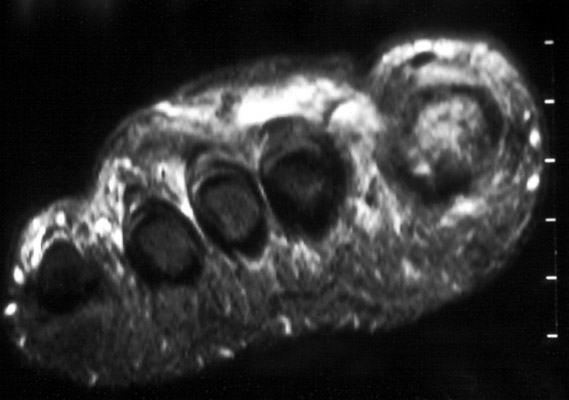
Sinus Tracts Sinus tracts are best demonstrated on MR imaging. Small tracts, however, may be difficult to find. To improve sensitivity, the radiologist should be aware of where drainage occurs to the skin. Markers placed at these sites help to focus the examination. T1 weighted images with intravenous gadolinium may be more sensitive in finding smaller abscesses and sinus tracts.
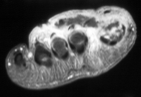
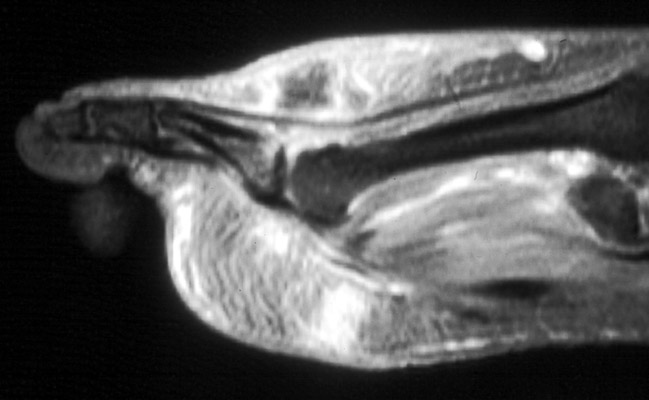
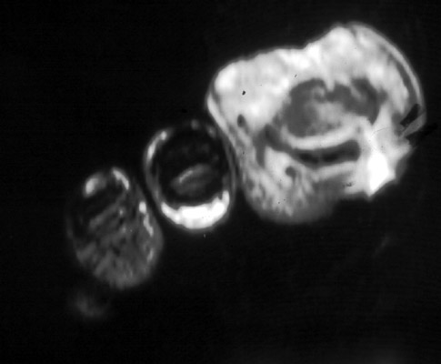
Shorts axis STIR and T1 weighted images with fat saturation post intravenous gadolinium both demonstrate abscess in the dorsum of the foot with associated sinus tract. The sagittal T1 weighted images with fat saturation post intravenous gadolinium demonstrates the abscess as well as underlying osteomyelitis of the proximal phalange with disruption of the proximal superior bone cortex. (Click on the images to see larger versions)
|
|
Advanced Imaging of the Diabetic Foot and Its
Complications
>
|