![]()
![]()
![]()
![]()
![]()
![]()
![]()
![]()
![]()
![]()
![]()
![]()
![]()
|
|
|
t
t
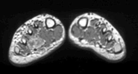
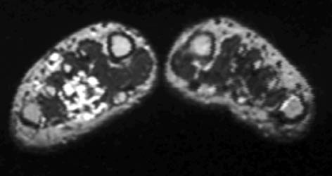
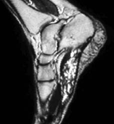
HemangiomaTypically divided into capillary (containing small capillary-like vessels) and cavernous (containing large sinusoids filled with blood) categories. Although lesions are most common in the skin and subcutaneous tissues, they also involve the muscles of the peripheral extremities. They are often irregular and have mixed signal intensity. Usually they are slightly higher in intensity than skeletal muscle on T1 and have markedly high intensity on T2 weighted sequences, with internal septa and slightly lobulated or irregular borders. Numerous serpiginous vessels may be seen.
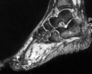
39 year old male with an extensive soft tissue mass involving the plantar aspect of the foot, dissecting between the third and fourth metatarsals, and extending into dorsum of the foot. The mass has intermediate signal on T1 and very high signal on T2 weighted images, and appears serpiginous. |
|
|