![]()
![]()
![]()
![]()
![]()
![]()
![]()
![]()
![]()
![]()
![]()
![]()
![]()
|
|
|
t
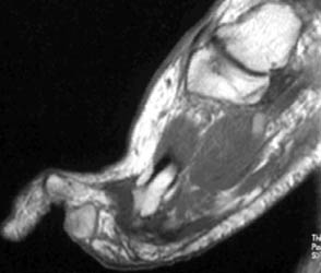
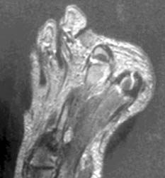
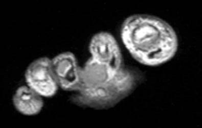
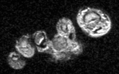
Metastatic Malignant MelanomaMalignant melanoma develops from melanocytes derived from neural crest cells, usually occurring in the skin. Other primary sites include the scalp, oral and anogenital mucosal surfaces, nail beds, conjunctivae, orbit, esophagus, and leptomeninges. Metastases occur after a latent period of 2 to 20 years in lymph nodes, bone, lung, liver, spleen , GI tract, kidneys, adrenals, and subcutaneous tissue. Nonhemorrhagic melanotic metastases have high signal on T1 and intermediate to low signal on T2 W images, caused by intrinsic paramagnetic effects. These may be due to paramagnetic cations, free radicals, or an inherent characteristic of melanin. However, amelanotic melanoma without hemorrhage may have low signal on T1 and high signal on T2W images.
63-year-old male with a 1.2 x 0.9 cm mass in the plantar soft tissues between the 2nd and 3rd proximal phalanges. This mass has high signal on T1, intermediate signal on proton density, and low signal on T2 W images, consistent with melanotic metastatic melanoma. |
|
|