|
Up
Wound Infection
Seroma and Epidural Scar
Epidural Hematoma - CT
Epidural Hematoma - MRI
Instability
Graft Reabsorption
Endplate Fracture
Graft Collapse
Nonunion
Displaced Strut Graft
Broken Screw
Screw in Spinal Canal
Slipped rod
Screw in Disc space
Screw Loosening & Hematoma
Screw Backing Out
Screw Loosening
| |
Epidural Hematoma
75 y.o. female with about a two-year history of continued gait abnormalities
and decreased dexterity. On examination her strength was normal, however, she
had bilateral Hoffmann signs as well as bilateral Babinski signs. Her strength
was 5/5 but her gait was ataxic. Magnetic resonance imaging showed severe
stenosis at C3, C5 and C6 levels. She underwent C3-C6 laminectomies.
Immediately post-operatively she did well, however, within 36 hours of the
operation, she slipped and fell due to spilled milk on the floor . After that
injury, the patient was neurologically intact. However, on postoperative day #2
for morning rounds, the patient was noted to be confused and weakened on the
left side, both arm and leg, leg more than the arm. An immediate CAT scan of the
brain and cervical spine was obtained. Immediate MRI was also obtained.
|
 A A
 B B
 C C
Click on the images to view a larger version!
|
|
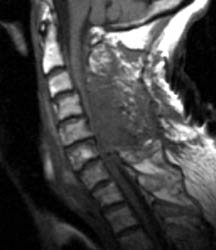
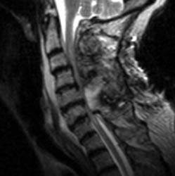
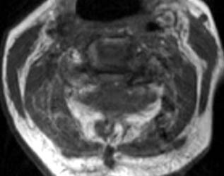
Non-enhanced sagittal T1 (A), sagittal T2 (B) and axial T1
images demonstrate a large fluid collection,
predominantly low signal on T1, bright on T2 with a few areas of T1 hyperintesity
mixed within. These constellation of findings are consistent with a
post-operative epidural hematoma with blood products of various ages. There is
severe severe spinal canal stenosis at C3-4, and very severe spinal canal
stenosis at C4-5 and C5-6. The cord is markedly flattened in the
anterior-posterior dimension, to approximately 2-3 mm secondary to mass effect
from this fluid collection. There is abnormal T2 weighted hyperintensity within
the substance of the cord at the C5-6 and C6-7 levels, consistent with some
edema.
|
|