Infection
Home Up Materials Radiography Arthrography CT and MRI References


Home
Materials
Fixation
Prosthesis Types
 Total Hip 
 Hemi
 Revision 
Radiography
Baseline Exam
 Alignment
 -Acetab. Position
 -Inclination
 -Anteversion
 -Center of Rotation
 -Femoral Stem 
 -Leg Length Discr.
 Intra-op- Fractures 
 Cement Herniation
Follow-up Study
 Bony Abnormalities
 Fracture
 Heterotopic Bone
 Non Union
 Hardware Failure
 -Polyethylene Wear
 -Dislocation
 Osteolysis
 Loosening
 Infection
Arthrography
CT and MRI
References

INFECTION

  • Incidence
    • 1 to 2 per cent of primary arthroplasties and 3 to 4 per cent of revisions.[1,2,3,4,5,6,7]
  • Radiographic findings
    • Similar to findings in loosening with progressive interface widening
    • There are no definitive radiographic signs to differentiate loosening from infection except for soft tissue gas
    • Soft tissue gas (ulcer or sinus tract)
    • Laminated periosteal reaction (rare)
  • Diagnostic evaluation
    • Blood tests
    • Aspiration – arthrography
    • Nuclear medicine
  • Treatment
    • Acute e.g. after dental surgery
      • Open surgical lavage
    • Sub acute or chronic
      • Resection of hardware with flail hip (Girdlestone)
      • Resection of hardware and placement of cement spacer

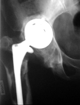
INFECTION—Markedly widened interfaces about acetabular and femoral components


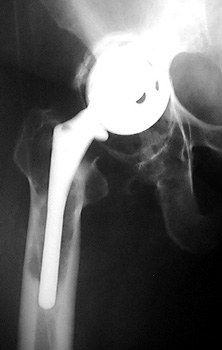
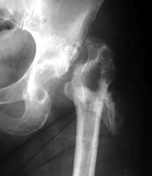
INFECTION Progressive interface widening about acetabular component


INFECTION—Soft tissue gas. 

Gas in neo capsule of right total hip replacement secondary to sinus tract from joint to skin


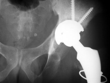
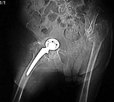
INFECTION—Soft tissue gas adjacent to dislocated cement spacer


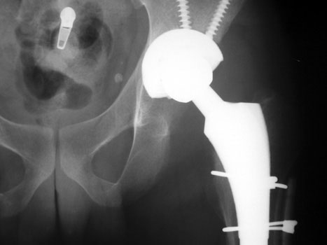
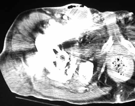
INFECTION—Gas bubbles in synovial cyst anterior to right total hip replacement. Patient had infected left hip joint 1 year previously treated by Girdlestone procedure. Right total hip replacement was grossly loose and with migrated hardware.


INFECTION
Resection of hardware, flail hip


INFECTION
Resection of hardware, cement spacer placement


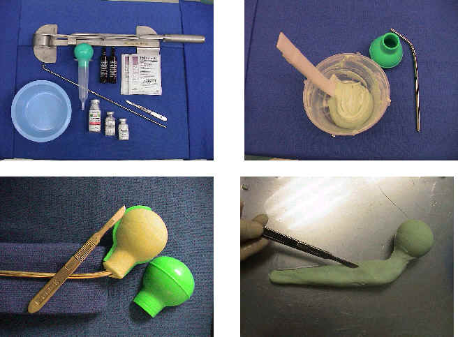
Cement spacers are antibiotic impregnated, allowing for local dispersal of antibiotics.

  • Maintain leg length
  • Minimize dead space
  • Preserve soft tissue planes
  • Facilitate ease of revision arthroplasty

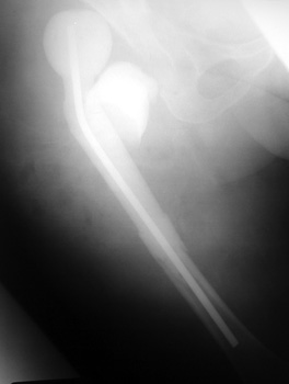
Cement spacers are fabricated in the surgical suite using methylmethacralate cement impregnated with antibiotics sensitive to cultured organisms. A Rush rod is cut and bent to 120 degrees to mimic the femoral neck-shaft angle. Antibiotic impregnated cement is placed into a blue bulb syringe which simulates the femoral head contour and allowed to dry. The plastic is then removed. A second batch of cement is prepared and formed around the femoral shaft portion of the Rush rod.


Google
 
Web Gentili.net